Human Leg Bone Diagram / Bones Of The Leg And The Foot Skeleton Of The Hindlimb Anatomy Americanhighschool Anatomylessons Ahsrocks A Anatomy Bones Leg Anatomy Human Body Anatomy / The nerves of the leg and foot arise from spinal nerves connected to the spinal cord in the lower back and pelvis.
byAdmin-
0
Human Leg Bone Diagram / Bones Of The Leg And The Foot Skeleton Of The Hindlimb Anatomy Americanhighschool Anatomylessons Ahsrocks A Anatomy Bones Leg Anatomy Human Body Anatomy / The nerves of the leg and foot arise from spinal nerves connected to the spinal cord in the lower back and pelvis.. The femur, or thigh bone, is the largest, heaviest, and strongest bone in the human body. The bones of the leg are the femur, tibia, fibula and patella. They support the legs to bear the body weight and also help in proper locomotion. Continue scrolling to read more below. The gastrocnemius is the larger calf muscle, forming the bulge visible beneath the skin.
Beside that, we also come with more related ideas as follows free printable human anatomy coloring pages, lower leg muscle diagram blank and lower limb bones unlabeled. Long bones are found in the arms (humerus, ulna, radius) and legs (femur, tibia, fibula), as well as in. The pubis, ischium, and ilium together constitute the pelvis while the thigh bone is the femur. The lumbar plexus forms in the lower back from the merger of spinal nerves l1 through l4 while the. Anatomy, medicine and biology concept.
1 701 Human Femur Bone Photos Free Royalty Free Stock Photos From Dreamstime from thumbs.dreamstime.com Posted on june 8, 2016 by admin. The skeletal system the human skeletal system, vector illustrations of human skeleton front and rear view leg bone stock illustrations. The lower leg is comprised of two bones, the tibia and the smaller fibula. The bones of the leg are the femur, tibia, fibula and patella.the foot bones shown in this diagram are the talus, navicular, cuneiform, cuboid, metatarsals and … as these muscles contract and relax, they move skeletal … Our goal is that these leg anatomy worksheets pictures gallery can be a direction for you, bring you more references and also make you have a great day. Long bones are found in the arms (humerus, ulna, radius) and legs (femur, tibia, fibula), as well as in. The knee joint is the largest joint in the body and is primarily a hinge joint, although some sliding and rotation occur. Continue scrolling to read more below.
The thigh bone, or femur, is the large upper leg bone that connects the lower leg bones (knee joint) to the pelvic bone (hip joint).
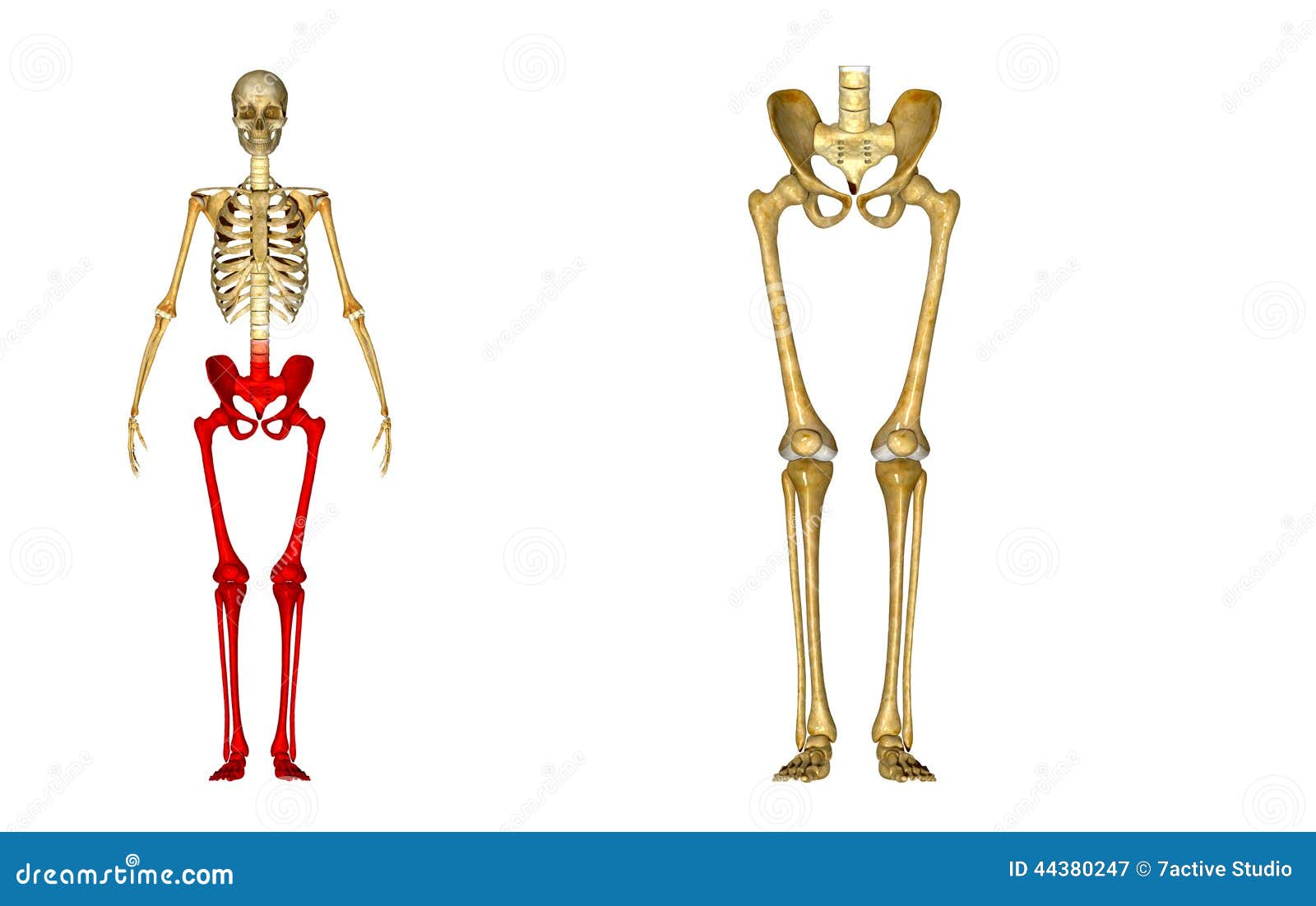
The hip itself is a ball and socket joint, much like the shoulder.the structures necessary to create this joint are the socket, the joint capsule, muscle, ligaments, and the neck. The hip itself is a ball and socket. The nerves of the leg and foot arise from spinal nerves connected to the spinal cord in the lower back and pelvis. The calf muscle, on the back of the lower leg, is actually made up of two muscles: This diagram depicts leg parts anatomy with parts and labels. Femur, upper bone of the leg or hind leg. He leg's main function in the human is for locomotion and support of the rest of the body. The lower leg is comprised of two bones, the tibia and the smaller fibula. The foot bones shown in this diagram are the talus, navicular, cuneiform, cuboid, metatarsals and calcaneus. Human leg bone diagram.the human leg, in the common word sense, is the entire lower limb of the human body. 6 / 10 ( 2 votes ) muscle of the human leg diagram. Related posts of diagram of leg bones bone anatomy elbow. The bones of the leg are the femur, tibia, fibula and patella.the foot bones shown in this diagram are the talus, navicular, cuneiform, cuboid, metatarsals and calcaneus.
Femur, upper bone of the leg or hind leg. The thigh bone, or femur, is the large upper leg bone that connects the lower leg bones (knee joint) to the pelvic bone (hip joint). Leg bone anatomy diagram diagram of human leg human anatomy diagram 10 / 10 ( 1 vote ) in this image, you will find femur, medial epicondyle of the femur, patella, tibial tuberosity, anterior rest of the tibia, a medial surface of the tibia, lateral epicondyle of the femur, head of the fibula, fibula, medial malleolus of the tibia, lateral. This diagram depicts leg parts anatomy with parts and labels. He leg's main function in the human is for locomotion and support of the rest of the body.
Bone Human Anatomy Knee Human Leg Crus Chinese Bones Angle Hand Foot Png Pngwing from w7.pngwing.com Related posts of muscles and tendons of the leg muscle anatomy diagram. Vector illustration isolated on a dark grey background useful for creating medical and scientific materials. Related posts of diagram of leg bones bone anatomy elbow. The ilium is the big bone of the hip, the ischium is the bone on which one sits and the pubis forms the lower frontal hip bone as seen in the diagram. He leg's main function in the human is for locomotion and support of the rest of the body. The lumbar plexus forms in the lower back from the merger of spinal nerves l1 through l4 while the. This diagram depicts leg parts anatomy with parts and labels. The hip itself is a ball and socket.
Posted in diagrams circulation system.
The nerves of the leg and foot arise from spinal nerves connected to the spinal cord in the lower back and pelvis. Browse 7,053 leg bone stock photos and images available, or search for human leg bone or leg bone xray to find more great stock photos and pictures. Check out the diagram of leg muscle anatomy in the first diagram below. Our goal is that these leg anatomy worksheets pictures gallery can be a direction for you, bring you more references and also make you have a great day. He leg's main function in the human is for locomotion and support of the rest of the body. Beside that, we also come with more related ideas as follows free printable human anatomy coloring pages, lower leg muscle diagram blank and lower limb bones unlabeled. In the leg muscles diagram above, there are many muscles that make up your legs and support it to move. The lumbar plexus forms in the lower back from the merger of spinal nerves l1 through l4 while the. The pubis, ischium, and ilium together constitute the pelvis while the thigh bone is the femur. The knee joint is the largest joint in the body and is primarily a hinge joint, although some sliding and rotation occur. The thigh bone, or femur, is the large upper leg bone that connects the lower leg bones (knee joint) to the pelvic bone (hip joint). Tendons connect the knee bones to the leg muscles that move the knee. This diagram depicts leg parts anatomy with parts and labels.
These leg muscle diagrams show you the major muscles of the human leg. The skeletal system the human skeletal system, vector illustrations of human skeleton front and rear view leg bone stock illustrations. Tendons connect the knee bones to the leg muscles that move the knee. Any disorder or defect in the knee bone can restrict the activities of the leg which can directly affect our locomotion. Human leg bone diagram.the human leg, in the common word sense, is the entire lower limb of the human body.
Ortho Blog The Human Skeletal System How You Move from 2cjaxfltpa-flywheel.netdna-ssl.com The femur, or thighbone, is the longest and largest bone in the human body. The longest and the strongest bone in the human skeletal system as you can observe in the labeled skeleton diagram of the human body. The bones of the leg are the femur, tibia, fibula and patella.the foot bones shown in this diagram are the talus, navicular, cuneiform, cuboid, metatarsals and … as these muscles contract and relax, they move skeletal … The bones of the leg are the femur, tibia, fibula and patella.the foot bones shown in this diagram are the talus, navicular, cuneiform, cuboid, metatarsals and calcaneus. In humans the neck of the femur connects the shaft and head at a 125 degree angle, which is efficient for walking. The gastrocnemius is the larger calf muscle, forming the bulge visible beneath the skin. Its lower end helps create the knee joint. Anatomy, medicine and biology concept.
The lumbar plexus forms in the lower back from the merger of spinal nerves l1 through l4 while the.
The bones together make up the hip. The hip itself is a ball and socket. The knee joint is the largest joint in the body and is primarily a hinge joint, although some sliding and rotation occur. This area is commonly referred to as the calf. 6 / 10 ( 2 votes ) muscle of the human leg diagram. The femur, or thighbone, is the longest and largest bone in the human body. The femur or the thigh bone is closest to the body. The skeletal system the human skeletal system, vector illustrations of human skeleton front and rear view leg bone stock illustrations. The longest and the strongest bone in the human skeletal system as you can observe in the labeled skeleton diagram of the human body. The nerves of the leg and foot arise from spinal nerves connected to the spinal cord in the lower back and pelvis. In this image, you will find muscle of the human leg diagram, hip and femur middle layer, hip and femur deep layer, overview of the most important muscles of the leg, femur middle layer, femur deep layer, rectus femoris m. The lumbar plexus forms in the lower back from the merger of spinal nerves l1 through l4 while the. Legs are used for standing, and all forms of.
The bones together make up the hip leg bone diagram. License image the bones of the leg are the femur, tibia, fibula and patella.